Mitraclip Device (Clip) Application
- Home
- Mitraclip Device (Clip) Application
Mitraclip Device (Clip) Application
This device is used especially in patients with mitral insufficiency who are unable to undergo surgery. The mitral valve, located between the left ventricle and the left atrium, should close during the contraction of the left ventricle and prevent blood from flowing back into the left atrium. However, in cases of severe mitral insufficiency, significant regurgitation occurs. If this condition persists over time, it can lead to heart enlargement and a progressive deterioration of the left ventricle’s contraction function. In normal individuals, the ejection fraction (ejection rate) reflecting the contraction of the left ventricle is between 55-75%, but when this rate falls below 30-35%, open heart surgery to replace the mitral valve is no longer possible. In this case, the only option is mitral valve replacement.
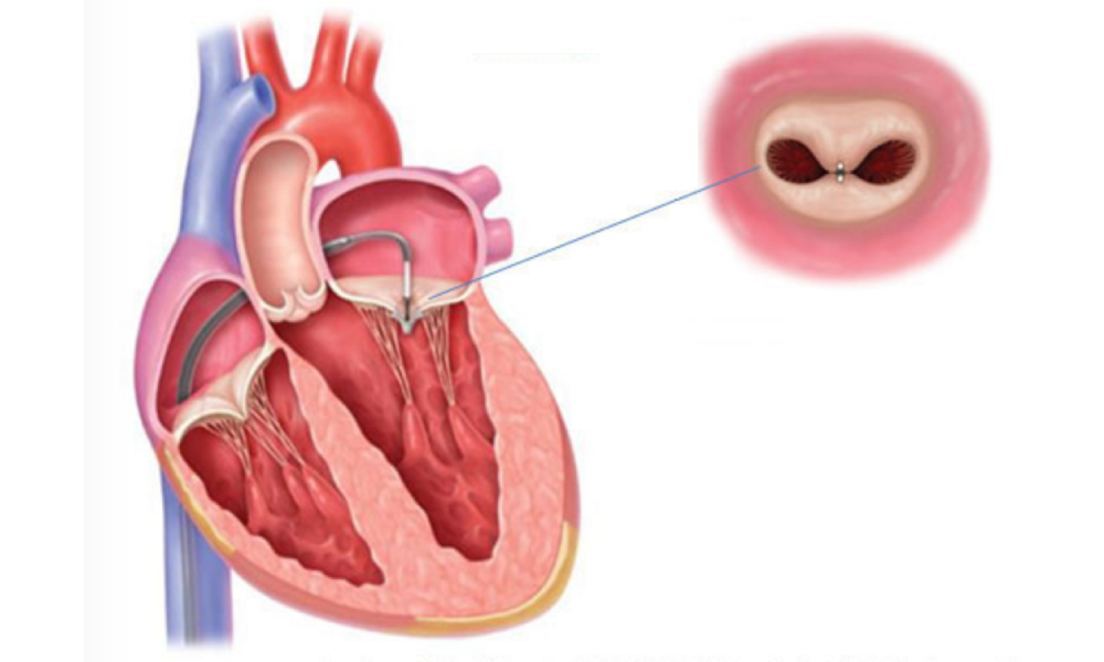
During this procedure, a needle is inserted into the right or left groin (femoral vein) and the septum between the right and left atria is punctured with a needle to reach the left atrium. A catheter (plastic tube) is used to deliver the MitraClip device from the right atrium to the left atrium. The clip-shaped device is attached to the front and back leaflets of the mitral valve under the guidance of TEE (transoesophageal echocardiography).
In this way, without the need for open heart surgery, i.e. without stopping the heart, connecting it to a pump, or opening the chest, the mitral valve, which is not completely closed and allows blood to leak into the left atrium, is repaired without surgery.
Patients with advanced mitral insufficiency who are at very high surgical risk and cannot undergo surgery can now be easily treated with this method. After placing the clip, two openings are created in the valve, whereas normally there is only one.
The valve contracts, reducing the amount of blood flowing into the left atrium. In some cases, multiple MitraClips may be required. If the procedure is successful, the left atrium’s contraction strength increases, symptoms of heart failure decrease, the patient’s quality of life improves, and life expectancy is extended.
 Türkçe